

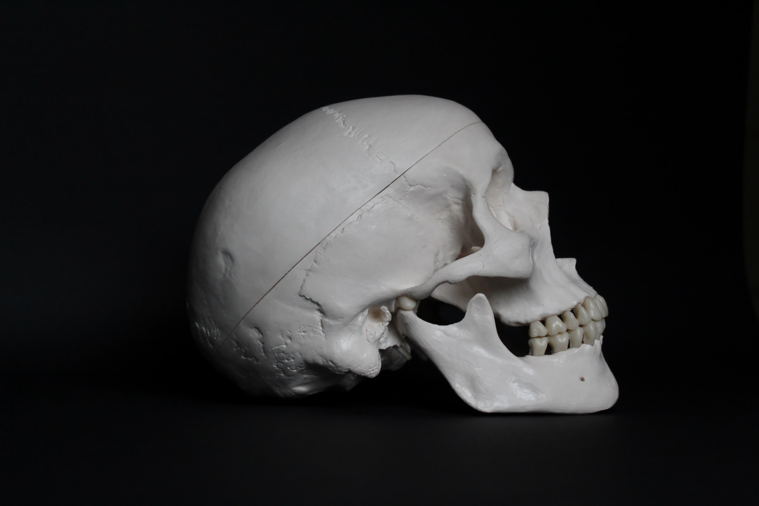
How poor oral hygiene may result in metabolic syndrome
Researchers have identified a novel mechanism by which periodontal disease may cause metabolic syndrome. By studying patients with metabolic syndrome, the researchers demonstrated high antibody titers against Porphyromonas gingivalis, the bacterium causing periodontal disease. In a mouse model, the researchers then showed that infection with this bacterium causes systemic insulin resistance and metabolic dysfunction in skeletal muscle by altering the gut microbiome. This study shows the effect periodontal disease can have on the entire body.
Periodontal or gum disease is known to be a significant risk factor of metabolic syndrome, a group of conditions increasing the risk for heart disease and diabetes. In a new study, researchers from Tokyo Medical and Dental University (TMDU) discovered that infection with Porphyromonas gingivalis, the bacterium causing periodontal disease, causes skeletal muscle metabolic dysfunction, the precursor to metabolic syndrome, by altering the composition of the gut microbiome.
Periodontal bacteria have long been known to cause inflammation within the oral cavity, but also systemically increase inflammatory mediators. As a result, sustained infection with periodontal bacteria can lead to increases in body weight and lead to increased insulin resistance, a hallmark of type 2 diabetes. The function of insulin is to help shuttle glucose from the blood into tissues, most importantly to skeletal muscle, where one quarter of all glucose in stored. Unsurprisingly, insulin resistance plays a key role in the development of metabolic syndrome, a group of conditions including obesity, altered lipid metabolism, high blood pressure, high blood glucose levels, and systemic inflammation. Although skeletal muscle plays a key role in decreasing blood glucose levels, a direct connection between periodontal bacterial infection and the metabolic function of skeletal muscle has not been established yet.
"Metabolic syndrome has become a widespread health problem in the developed world," says first author of the study Kazuki Watanabe. "The goal of our study was to investigate how periodontal bacterial infection might lead to metabolic alterations in skeletal muscle and thus to the development of metabolic syndrome."

A tiny jaw from Greenland sheds light on the origin of complex teeth
Scientists have described the earliest known example of dentary bone with two rows of cusps on molars and double-rooted teeth. The new findings offer insight into mammal tooth evolution, particularly the development of double-rooted teeth.
A team of scientists led from Uppsala University have described the earliest known example of dentary bone with two rows of cusps on molars and double-rooted teeth. The new findings offer insight into mammal tooth evolution, particularly the development of double-rooted teeth. The results are published in the scientific journal PNAS.
The first mammals originated in the latest Triassic period, around 205 million years ago. An ancestor to mammals were the therapsids, "mammal-like reptiles" referred to as stem mammals or proto-mammals, which originated about 320-300 million years ago. One unique characteristic of the lineage that included mammals and animals related to mammals (synapsids) was that they developed complex occlusion. Close ancestors to mammals, called mammaliaforms, developed rows of cusps on molar-like teeth adapted for more omnivorous feeding. The origin of this multicusped pattern and double-rooted tooth has thus far remained unclear.
A team of scientists led by Grzegorz Niedzwiedzki from Uppsala University have investigated the jaw anatomy and tooth structure of a recently described new mammaliaform species named Kalaallitkigun jenkinsi. It was discovered on the eastern coast of Greenland and was a very small, shrew-like animal, probably covered with fur. It would have been the size of a large mouse and lived during the Late Triassic, around 215 million years ago.

Researchers ask: how sustainable is your toothbrush?
Researchers have examined the sustainability of different models of the most commonly used oral health product - the toothbrush - to ascertain which is best for the planet and associated human health.
Researchers at Trinity College Dublin have examined the sustainability of different models of the most commonly used oral health product -- the toothbrush -- to ascertain which is best for the planet and associated human health.
Although the toothbrush is a widely recommended healthcare device worldwide, there is currently little quantitative data available for its impact on the planet. The research study, in collaboration with Eastman Dental Institute at University College London, is published in the British Dental Journal today (Tuesday, 15th September 2020). It represents the first time a life-cycle assessment (LCA) has been used to measure environmental consequences of a healthcare product.
Healthcare is a major emitter of environmental pollutants that adversely affect health, but awareness of these effects remains low both in the industry and in the general consumer population. There is currently little evidence or guidance regarding the sustainability of specific healthcare interventions, services or devices

Gum disease may raise risk of some cancers
People with history of gum disease appear to have higher risk of developing oesophageal and gastric cancer, suggest researchers
People who have periodontal (gum) disease may have a higher risk of developing some forms of cancer, suggests a letter published in the journal Gut detailing a prospective study.
US researchers found that a history of periodontal disease appeared to be associated with a raised risk of esophageal (gullet) cancer and gastric (stomach) cancer and this risk was also higher among people who had lost teeth previously.
Previous findings on the relationship of periodontal disease and tooth loss with esophageal and gastric cancer have been inconsistent.
Therefore, a team of researchers from Harvard T.H. Chan School of Public Health, in Boston, USA, carried out a study of data on patients over decades of follow up.
They examined the association of history of periodontal disease and tooth loss with the risk of esophageal and gastric cancer in 98,459 women from the Nurses' Health Study (1992-2014) and 49,685 men from the Health Professionals Follow-up Study (1988-2016).
Dental measures, demographics, lifestyle, and diet were assessed using follow-up questionnaires and self-reported cancer diagnosis was confirmed after reviewing medical records.
The results showed that during 22-28 years of follow-up, there were 199 cases of esophageal cancer and 238 cases of gastric cancer.
A history of periodontal disease was associated with a 43% and 52% increased risk of esophageal cancer and gastric cancer, respectively.
Compared to people with no tooth loss, the risks of esophageal and gastric cancer for those who lost two or more teeth were also modestly higher -- 42% and 33%, respectively.
In addition, among individuals with a history of periodontal disease, no tooth loss and losing one or more teeth were equally associated with a 59% increased risk of esophageal cancer compared to those with no history of periodontal disease and no tooth loss.
Similarly, the same group of individuals had 50% and 68% greater risk of gastric cancer, respectively.

Her Song Donations
CJ Henley, DMD is partnering with NEFDHA (Northeast Florida Dental Hygienists Association) in collecting donations for Her Song Jacksonville. Her Song Jacksonville is where young ladies from Jacksonville and all over the nation find refuge in the aftermath of human trafficking
CJ Henley, DMD is partnering with NEFDHA (Northeast Florida Dental Hygienists Association) in collecting donations for Her Song Jacksonville. Her Song Jacksonville is where young ladies from Jacksonville and all over the nation find refuge in the aftermath of human trafficking. It provides residential safe homes with comprehensive programs in the Northeast Florida area for sex trafficking victims. The program assists young women in exiting cycles of abuse and trauma and empower the exploited to heal and take back their lives. Her Song has worked with more than 800 survivors of human trafficking since its inception. We are working to give these courageous young woman a memorable Christmas this season.
Please see attached wish lists.
Cleaning supply list that are needed
Clorox Wipes
Bleach
Windex
Lysol Spray
Lysol wipes
All-purpose cleaning spray
Dish detergent
Dish washer detergent
Paper Towels
Sponges
Scrubbers
Brillo Pads
Toilet bowl cleaner
Bathroom/tub cleaner
Toilet Paper
Air Freshener Spray
Hand Soap
Laundry Pods
Dryer Sheets
Fabric Softener
Swiffer WetJet
Cleaning gloves
https://www.amazon.com/hz/wishlist/ls/1FYMVD460ZWOY?ref_=wl_sh
A tiny jaw from Greenland sheds light on the origin of complex teeth
Scientists have described the earliest known example of dentary bone with two rows of cusps on molars and double-rooted teeth. The new findings offer insight into mammal tooth evolution, particularly the development of double-rooted teeth.
A team of scientists led from Uppsala University have described the earliest known example of dentary bone with two rows of cusps on molars and double-rooted teeth. The new findings offer insight into mammal tooth evolution, particularly the development of double-rooted teeth. The results are published in the scientific journal PNAS.
The first mammals originated in the latest Triassic period, around 205 million years ago. An ancestor to mammals were the therapsids, "mammal-like reptiles" referred to as stem mammals or proto-mammals, which originated about 320-300 million years ago. One unique characteristic of the lineage that included mammals and animals related to mammals (synapsids) was that they developed complex occlusion. Close ancestors to mammals, called mammaliaforms, developed rows of cusps on molar-like teeth adapted for more omnivorous feeding. The origin of this multicusped pattern and double-rooted tooth has thus far remained unclear.
A team of scientists led by Grzegorz Niedzwiedzki from Uppsala University have investigated the jaw anatomy and tooth structure of a recently described new mammaliaform species named Kalaallitkigun jenkinsi. It was discovered on the eastern coast of Greenland and was a very small, shrew-like animal, probably covered with fur. It would have been the size of a large mouse and lived during the Late Triassic, around 215 million years ago.
"I knew it was important from the moment I took this 20 mm specimen off the ground," says Niedzwiedzki, researcher at Uppsala University and the corresponding author of the publication.
Kalaallitkigun jenkinsi exhibits the earliest known dentary with two rows of cusps on molars and double-rooted teeth. The anatomical features place Kalaallitkigun jenkinsi as an intermediate between the mammals and the insectivorous morganucodontans, another type of mammaliaform.
The researchers believe that the structural changes in the teeth are related to changed feeding habits. In this case study, the animals were switching to a more omnivorous/herbivorous diet and the tooth crown was expanding laterally. Broader teeth with "basins" on the top surface are better for grinding food. This development also forced changes in the structure of the base of the tooth.
The biomechanical analysis that was carried out within the study found that multi-rooted teeth are better able to withstand mechanical stresses, including those of upper and lower tooth contact during biting, compared to single-rooted teeth. Human teeth, for instance, have this characteristic. The results suggest that the development of molar-like teeth with complex crowns may have developed together with biomechanically optimised dual roots.
"The early evolution of mammals is a particularly interesting topic in evolutionary studies. This tiny jaw from Greenland shows us how complex mammalian teeth arose and why they appeared," says Niedzwiedzki.
"Our discovery of the oldest mammalian ancestor with double-rooted molars shows how important the role of teeth was in the origin of mammals. I had this idea to look at the biomechanics and the collaboration with the engineers turned out great," says Tomasz Sulej, researcher at the Polish Academy of Sciences, first author of the publication.
"It seems that the fossils of close mammalian ancestors must be looked for in even older rocks," says Sulej.
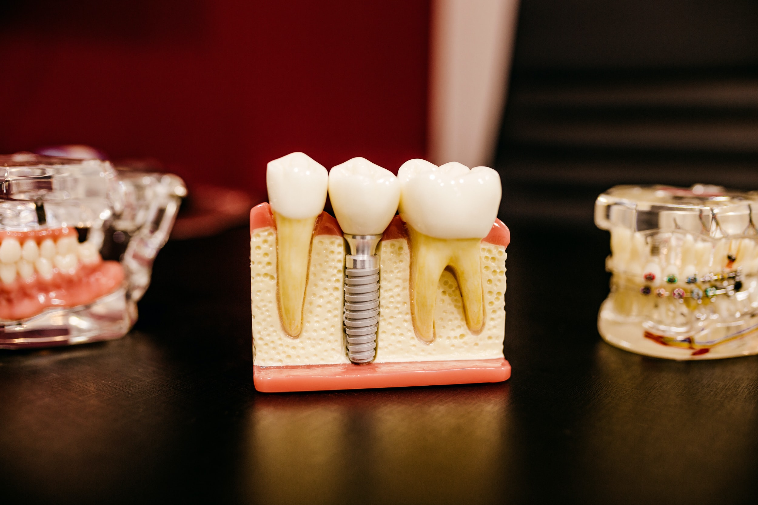
Improving the treatment of periodontitis
For the first time, researchers have shown that a unicellular parasite commonly found in the mouth plays a role in both severe tissue inflammation and tissue destruction.
For the first time, researchers from Charité -- Universitätsmedizin Berlin have shown that a unicellular parasite commonly found in the mouth plays a role in both severe tissue inflammation and tissue destruction. Most patients with severe and recurrent periodontitis (gum disease) showed an increased presence of the amoeba Entamoeba gingivalis inside their oral cavities. The effect of this amoeba is similar to that of Entamoeba histolytica, the parasite responsible for causing amebiasis. Once the parasite has invaded the gingival tissue, it feeds on its cells and causes tissue destruction. According to the researchers' findings, which have been published in the Journal of Dental Research, the two amoebae show similar mechanisms of tissue invasion and elicit a similar immune response in the host.
Periodontitis, or gum disease, is an inflammation of the gums and supporting structures of the teeth. It is one of the most common chronic diseases in the world. In Germany, approximately 15 percent of people are affected by a particularly severe form of this disease. If left untreated, periodontitis will lead to tooth loss. The disease also increases the risk of arthritis, cardiovascular disease and cancer. In patients with periodontitis, a decrease in the diversity of the oral flora coincides with an increase in the frequency of E. gingivalis. A team of researchers, led by Prof. Dr. Arne Schäfer, Head of the Periodontology Research Unit at Charité's Institute of Dental and Craniofacial Sciences, was able to show that oral inflammation is associated with colonization by the oral parasite E. gingivalis.
Scientists have long been aware of the virulence potential of this genus of amoebae. The gastrointestinal parasite E. histolytica, for instance, causes a disease known as amebiasis, one of the most common causes of death from parasitic diseases worldwide. "We have shown that an amoeba like E. gingivalis, which colonizes the oral cavity, will invade the oral mucosa and destroy gingival tissue. This enables increased numbers of bacteria to invade the host tissue, which further exacerbates inflammation and tissue destruction," says Prof. Schäfer. The international team of researchers was the first to describe precise roles of E. gingivalis in the pathogenesis of inflammation. During their analysis of inflamed periodontal pockets, the researchers detected evidence of the amoeba in approximately 80 percent of patients with periodontitis, but in only 15 percent of healthy subjects. Their observations revealed that, after invading the gums, the parasites move within the tissue, feeding on and killing host cells. Cell culture experiments showed that infection with E. gingivalis slows the rate at which cells grow, eventually leading to cell death.
The researchers concluded that the amoeba's role in inflammation shows distinct parallels to the pathogenesis of amebiasis. "E. gingivalis actively contributes to cell destruction inside the gingival tissue and stimulates the same host immune response mechanisms as E. histolytica during its invasion of the intestinal mucosa," explains Prof. Schäfer. "This parasite, which is transmitted by simple droplet infection, is one potential cause of severe oral inflammation."
Treatment success is often short-lived in patients with periodontitis. This might be due to the high virulence potential of this previously unnoticed, yet extremely common amoeba. Summing up the results of the research, Prof. Schäfer says: "We identified one infectious parasite whose elimination could improve treatment effectiveness and long-term outcomes in patients with gum disease." He adds: "Current treatment concepts for periodontitis fail to consider the possibility of infection by this parasite or its successful elimination." A clinical trial is underway to determine the extent to which the elimination of this amoeba might improve treatment outcomes in patients with periodontitis.

Ouch: Patients prescribed opioids after tooth extraction report worse pain
The use of opioids to soothe the pain of a pulled tooth could be drastically reduced or eliminated altogether from dentistry, say researchers.
The use of opioids to soothe the pain of a pulled tooth could be drastically reduced or eliminated altogether from dentistry, say University of Michigan researchers.
More than 325 dental patients who had teeth pulled were asked to rate their pain and satisfaction within six months of extraction. Roughly half of the study's patients who had surgical extraction and 39% who had routine extraction were prescribed opioids.
The U-M researchers compared the pain and satisfaction of those who used opioids to those who didn't.
"I feel like the most important finding is that patient satisfaction with pain management was no different between the opioid group and non-opioid group, and it didn't make a difference whether it was surgical or routine extraction," said study co-author Romesh Nalliah, clinical professor and associate dean for patient services at the U-M School of Dentistry.
Surprisingly, patients in the opioid group actually reported worse pain than the non-opioid group for both types of extractions, Nalliah said.
The researchers also found that roughly half of the opioids prescribed remained unused in both surgical and nonsurgical extractions. This could put patients or their loved ones at risk of future misuse of opioids if leftover pills are not disposed of properly.
The findings are scheduled to appear March 13 in JAMA Network Open.
"The real-world data from this study reinforces the previously published randomized-controlled trials showing opioids are no better than acetaminophen and nonsteroidal anti-inflammatory drugs for pain after dental extraction," said study co-author Chad Brummett, director of the Division of Pain Research and of Clinical Research in the Department of Anesthesiology at Michigan Medicine, U-M's academic medical center.
Brummett co-directs the Michigan Opioid Prescribing Engagement Network, or Michigan OPEN, which has developed, tested and shared guidelines for the use of opioids in patients with acute pain from surgery and medical procedures.
"These data support the Michigan OPEN prescribing recommendations calling for no opioids for the majority of patients after dental extractions, including wisdom teeth extraction," he said.
The results have big implications for both patients and dentists, and suggest prescribing practices need an overhaul, Brummett and Nalliah said.
The American Dental Association suggests limiting opioid prescribing to seven days' supply, but Nalliah believes that's too high.
"I think we can almost eliminate opioid prescribing from dental practice. Of course, there are going to be some exceptions, like patients who can't tolerate nonsteroidal anti-inflammatories," he said. "I would estimate we can reduce opioid prescribing to about 10% of what we currently prescribe as a profession."
For dentists, many of whom are sole proprietors, this new information means they needn't worry so much about unhappy patients changing practices if they aren't prescribed strong opioids. Alternatives such as nonsteroidal anti-inflammatory drugs or acetaminophen appear to control pain better, and patient satisfaction remains high.
Nalliah gives two possible reasons for this. First, dentists may have prescribed opioids in only the toughest cases, which would have resulted in more pain regardless.
"Or alternatively, and this is the reason I tend to accept, is that our study concurs with previous studies that suggest opioids are not the most effective analgesic for acute dental pain," Nalliah said.
"Dentists are torn between wanting to satisfy patients and grow business and limiting their opioid prescribing in light of the current crisis. I think it's an extremely liberating finding for dentists who can worry more about the most effective pain relief rather than overprescribing for opioids."
Dentists account for about 6% to 6.5% of U.S. opioid prescriptions -- a relatively small amount. But the study notes that dentists are among the most common prescribers for minors, and for many patients, dental opioid prescriptions are their first exposure.

Could the cure for IBD be inside your mouth?
A new collaborative study reveals that inflammatory bowel disease (IBD) may be the latest condition made worse by poor oral health via a clash between the mouth and gut microbiomes.
A new collaborative study from the U-M Medical and Dental Schools reveals that inflammatory bowel disease (IBD), which included Crohn's disease and ulcerative colitis and afflicts an estimated 3 million adults in the U.S., may be the latest condition made worse by poor oral health.
Nobuhiko Kamada, Ph.D., assistant professor of internal medicine in the division of gastroenterology, has been studying the gut microbiome -- the collection of bacteria that are normally present in the gut -- for years. He noted an emerging link in research literature between an overgrowth of foreign bacterial species in the guts of people with IBD -- bacteria that are normally found in the mouth. "I decided to approach the dental school to ask the question, does oral disease affect the severity of gastrointestinal diseases?" says Kamada.
The new mouse study, published in Cell, shows two pathways by which oral bacteria appear to worsen gut inflammation.
In the first pathway, periodontitis, the scientific name for gum disease, leads to an imbalance in the normal healthy microbiome found in the mouth, with an increase of bacteria that cause inflammation. These disease-causing bacteria then travel to the gut.
However, this alone may not be enough to set off gut inflammation. The team demonstrated that oral bacteria may aggravate gut inflammation by looking at microbiome changes in mice with inflamed colons.
"The normal gut microbiome resists colonization by exogenous, or foreign, bacteria," says Kamada. "However, in mice with IBD, the healthy gut bacteria are disrupted, weakening their ability to resist disease-causing bacteria from the mouth." The team found that mice with both oral and gut inflammation had significantly increased weight loss and more disease activity.
In the second proposed pathway, periodontitis activates the immune system's T cells in the mouth. These mouth T cells travel to the gut where they, too, exacerbate inflammation. The gut's normal microbiome is held in balance by the action of inflammatory and regulatory T cells that are fine-tuned to tolerate the resident bacteria. But, says Kamada, oral inflammation generates mostly inflammatory T cells that migrate to the gut, where they, removed from their normal environment, end up triggering the gut's immune response, worsening disease.
"This exacerbation of gut inflammation driven by oral organisms that migrate to the gut has important ramifications in emphasizing to patients the critical need to promote oral health as a part of total body health and wellbeing," says co-author William Giannobile, DDS, the William K and Mary Anne Najjar professor of dentistry and chair of the department of periodontics and oral medicine at the U-M School of Dentistry.
The study has implications for novel treatments for IBD, necessary because "far too many patients still fail medications, leading to reduced quality of life and eventual surgery," says study co-author Shrinivas Bishu, M.D., assistant professor of gastroenterology. "This study importantly implies that clinical outcomes in IBD may be improved by monitoring oral inflammation -- an intriguing concept."

Harnessing pickle power to promote dental health
A research team evaluated 14 different types of Sichuan pickles from southwest China. They extracted 54 different strains of Lactobacilli and found that one, L. plantarum K41, significantly reduced the incidence and severity of cavities.
Can a probiotic derived from Chinese pickles prevent cavities? That seems to be the case, according to a study by researchers at Ben-Gurion University of the Negev and Chengdu University in China.
Pickles are an integral part of the diet in the southwest of China. When fruits and vegetables are fermented, healthy bacteria break down the natural sugars. These bacteria, also known as probiotics, not only preserve foods but offer numerous benefits, including immune system regulation, stabilization of the intestinal microbiota, reducing cholesterol levels, and now inhibiting tooth decay.
According to the study published in Frontiers in Microbiology, a strain of Lactobacilli (L. plantarum K41) found in Sichuan pickles reduced S. mutans by 98.4%. Dental caries (cavities) are caused by Streptococcus mutans, (S. mutans) commonly found in the human oral cavity as plaque and is a significant contributor to tooth decay.
Prof. Ariel Kushmaro of the BGU Avram and Stella Goldstein-Goren Department of Biotechnology Engineering and the Chinese research team evaluated 14 different types of Sichuan pickles from southwest China. They extracted 54 different strains of Lactobacilli and found that one, L. plantarum K41, significantly reduced the incidence and severity of cavities. K41 was also highly tolerant of acids and salts, an additional benefit as a probiotic for harsh oral conditions. It also could have potential commercial value when added to dairy products.
According to Doug Seserman, chief executive officer of American Associates, Ben-Gurion University of the Negev based in New York City, "the researchers currently have no plans to evaluate Jewish deli pickles."