


The Gut-Mouth Connection: How Heartburn Medication Impacts Gum Health
Can PPIs protect your gums? Discover the latest research on how heartburn medication affects gum disease severity. Medically-informed dental care in Jacksonville.
Research from the University at Buffalo suggests a surprising link between digestive health and the stability of your gums. The study found that patients using Proton Pump Inhibitors (PPIs)—common medications for heartburn and acid reflux—exhibited significantly less severe symptoms of gum disease (periodontitis).
As a practice focused on Complex & Medically Involved Care, we closely monitor how your systemic medications influence your oral clinical outcomes.
The Data: Smaller "Pockets" and Stronger Foundations
The study analyzed over 1,000 patients and used probing depths (the space between the tooth and gum) as the primary measure of health. Healthy gums fit snugly against the teeth, while deep pockets indicate bone loss and active infection.
The findings were striking:
Severe Disease: Only 14% of teeth in PPI users had deep pockets (6mm+), compared to 24% in non-users.
Moderate Disease: PPI users also showed a significant reduction in 5mm pockets compared to those not taking the medication.
Why Does Heartburn Medicine Affect the Gums?
While more research is needed, scientists believe the link may lie in how PPIs alter two critical systems:
Bone Metabolism: PPIs may influence how the body maintains the alveolar bone that supports your teeth.
The Microbiome: These drugs can shift the balance of bacteria in both the gut and the mouth, potentially reducing the "relative abundance" of harmful, gram-negative bacteria that drive inflammation.
Medically-Informed Periodontal Care
At CJ Henley, DMD, we don't treat the mouth in isolation. Understanding your full medical history—including your use of PPIs or other systemic drugs—is a vital part of our unhurried, two-hour new patient evaluation.
By coordinating with your medical team and understanding these systemic links, we can design more predictable, long-lasting restorative treatments that respect your body's unique physiology.
Reviewed and Updated February 2026
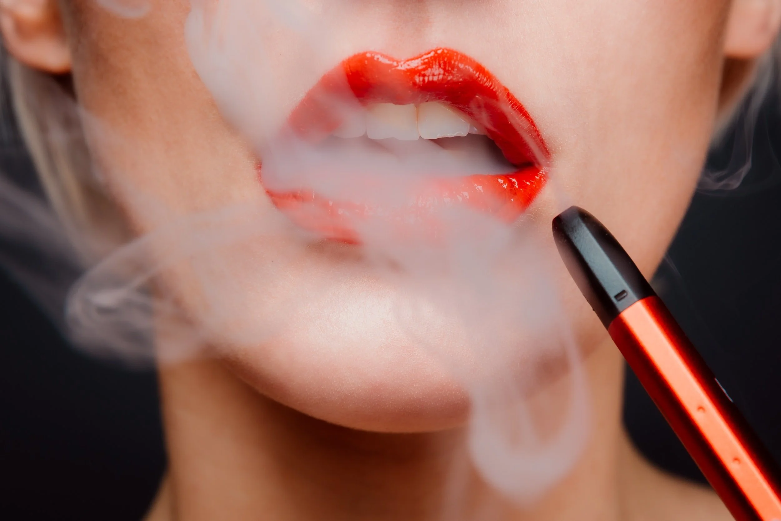
Evidence grows for vaping's role in gum disease
New studies highlight how e-cigarettes alter oral health and may be contributing to gum disease. The latest research finds that e-cigarette users have a unique oral microbiome that is less healthy than nonsmokers but potentially healthier than cigarette smokers, and measures worsening gum disease over time.
A series of new studies by researchers at NYU College of Dentistry highlights how e-cigarettes alter oral health and may be contributing to gum disease. The latest, published in mBio, finds that e-cigarette users have a unique oral microbiome -- the community of bacteria and other microorganisms -- that is less healthy than nonsmokers but potentially healthier than cigarette smokers, and measures worsening gum disease over time.
"To our knowledge, this is the first longitudinal study of oral health and e-cigarette use. We are now beginning to understand how e-cigarettes and the chemicals they contain are changing the oral microbiome and disrupting the balance of bacteria," said Deepak Saxena, who led the research with Xin Li; both are professors of molecular pathobiology at NYU College of Dentistry.
Gum disease affects nearly half of U.S. adults over 30 years of age. Smoking cigarettes is a known risk factor for developing gum disease, but less is known about the impact of e-cigarettes -- which vaporize nicotine and other chemicals -- on oral health, especially the long-term consequences of vaping.
The researchers studied the oral health of 84 adults from three groups: cigarette smokers, e-cigarette users, and people who have never smoked. Gum disease was assessed through two dental exams six months apart, during which plaque samples were taken to analyze the bacteria present.
Changes to gum health
All participants had some gum disease at the start of the study, with cigarette smokers having the most severe disease, followed by e-cigarette users. After six months, the researchers observed that gum disease had worsened in some participants in each group, including several e-cigarette users.
A key indicator of gum disease is clinical attachment loss, measured by gum ligament and tissue separating from a tooth's surface, leading the gum to recede and form pockets. These pockets are breeding grounds for bacteria and can lead to more severe gum disease. In a study of the same participants published in Frontiers in Oral Health, the research team found that clinical attachment loss was significantly worse only in the e-cigarette smokers -- not non-smokers and cigarette smokers -- after six months.
A unique microbiome
The researchers then analyzed the bacteria found in the plaque samples and determined that e-cigarette users have a different oral microbiome from smokers and nonsmokers -- building on findings the team previously reported in iScience and Molecular Oral Microbiology.
While all groups shared roughly a fifth of the types of bacteria, the bacterial makeup for e-cigarette users had strikingly more in common with cigarette smokers than nonsmokers. Several types of bacteria, including Selenomonas, Leptotrichia, and Saccharibacteria, were abundant in both smokers and vapers compared to nonsmokers. Several other bacteria -- including Fusobacterium and Bacteroidales, which are known to be associated with gum disease -- were particularly dominant in the mouths of e-cigarette users.
When plaque samples were gathered and analyzed in the six-month follow-up, the researchers found greater diversity in bacteria for all groups studied, yet each group maintained its own distinct microbiome.
"Vaping appears to be driving unique patterns in bacteria and influencing the growth of some bacteria in a manner akin to cigarette smoking, but with its own profile and risks to oral health," said Fangxi Xu, a junior research scientist in Saxena's lab and the study's co-first author.
An altered immune response
The researchers found that the distinct microbiome in e-cigarette users was correlated with clinical measures of gum disease and changes to the host immune environment. In particular, vaping was associated with different levels of cytokines -- proteins that help regulate the immune system. Certain cytokines are linked to an imbalance in oral bacteria and can worsen gum disease by making people prone to inflammation and infection.
TNFα, a cytokine that causes inflammation, was significantly elevated among e-cigarette users. In contrast, cytokines IL-4 and IL-1β were lower among e-cigarette users; IL-4 tends to be reduced in people with gum disease and increases after treatment, which suggests that certain bacteria in the mouths of e-cigarette users may be actively suppressing immune responses.
The researchers concluded that the distinct oral microbiome of e-cigarette users elicits altered immune responses, which along with clinical markers for gum disease illustrate how vaping presents its own challenge to oral health.
"E-cigarette use is a relatively new human habit," said Scott Thomas, an assistant research scientist in Saxena's lab and the study's co-first author. "Unlike smoking, which has been studied extensively for decades, we know little about the health consequences of e-cigarette use and are just starting to understand how the unique microbiome promoted by vaping impacts oral health and disease."
This research was supported by the National Institute of Dental & Craniofacial Research (DE025992, DE027074), the National Cancer Institute (CA206105), and the NYU Mega-Grants Initiative. The study included additional collaborators from NYU College of Dentistry, NYU Grossman School of Medicine, and the University of Pennsylvania.

Comparative Medicine: Why Oral Health is Vital for Every Family Member
Oral health impacts the heart, liver, and kidneys in both humans and pets. Discover the clinical links between periodontal disease and systemic wellness.
At CJ Henley, DMD, we focus on the specialized needs of Complex & Medically Involved Care in humans, but the biological principles of oral health extend across species. Periodontal disease is the most common dental condition in both humans and our animal companions, and the systemic consequences are remarkably similar.
Just as we coordinate with medical teams through the Baptist MD Anderson Head and Neck Tumor Board to protect human systemic health, veterinary professionals prioritize oral care to prevent damage to vital organs like the heart, liver, and kidneys.
The Systemic Link: More Than Just "Bad Breath"
In both humans and pets, periodontal disease starts with plaque that hardens into tartar. While tartar above the gumline is visible, the most significant damage occurs subgingivally (below the gumline). This hidden infection provides a direct gateway for bacteria to enter the bloodstream, potentially leading to:
Endocarditis: Inflammation of the heart muscle or valves.
Renal and Hepatic Stress: Chronic strain on the kidneys and liver as they filter oral bacteria from the blood.
Bone Loss: Irreversible damage to the jawbone and supporting structures.
The Clinical Standard: Why Professional Intervention Matters
Whether for a person or a pet, a "cleaning" is only effective if it addresses the pathology below the gumline. In veterinary medicine, this requires anesthesia to ensure a thorough, pain-free evaluation and to allow for diagnostic radiographs (X-rays) that reveal the health of tooth roots and the jaw.
In our practice, we utilize a similar philosophy of unhurried, thorough evaluation. We believe that early detection—identifying issues before they cause pain or systemic illness—is the highest standard of care.
Warning Signs of Oral Distress
While pets cannot communicate dental pain verbally, their clinical signs are often identical to the symptoms we treat in our Restorative Dentistry cases:
Discolored teeth or heavy tartar buildup.
Abnormal chewing or difficulty eating.
Swelling around the mouth or jaw.
Changes in behavior or irritability.
A Unified Approach to Wellness
Understanding the shared risks of oral infection helps us appreciate the importance of rigorous clinical standards. Whether we are managing Oncology-Related Dental Care for a human patient or discussing the needs of a family pet, the goal remains the same: protecting the integrity of the body through advanced oral medicine.
Reviewed and Updated February 2026
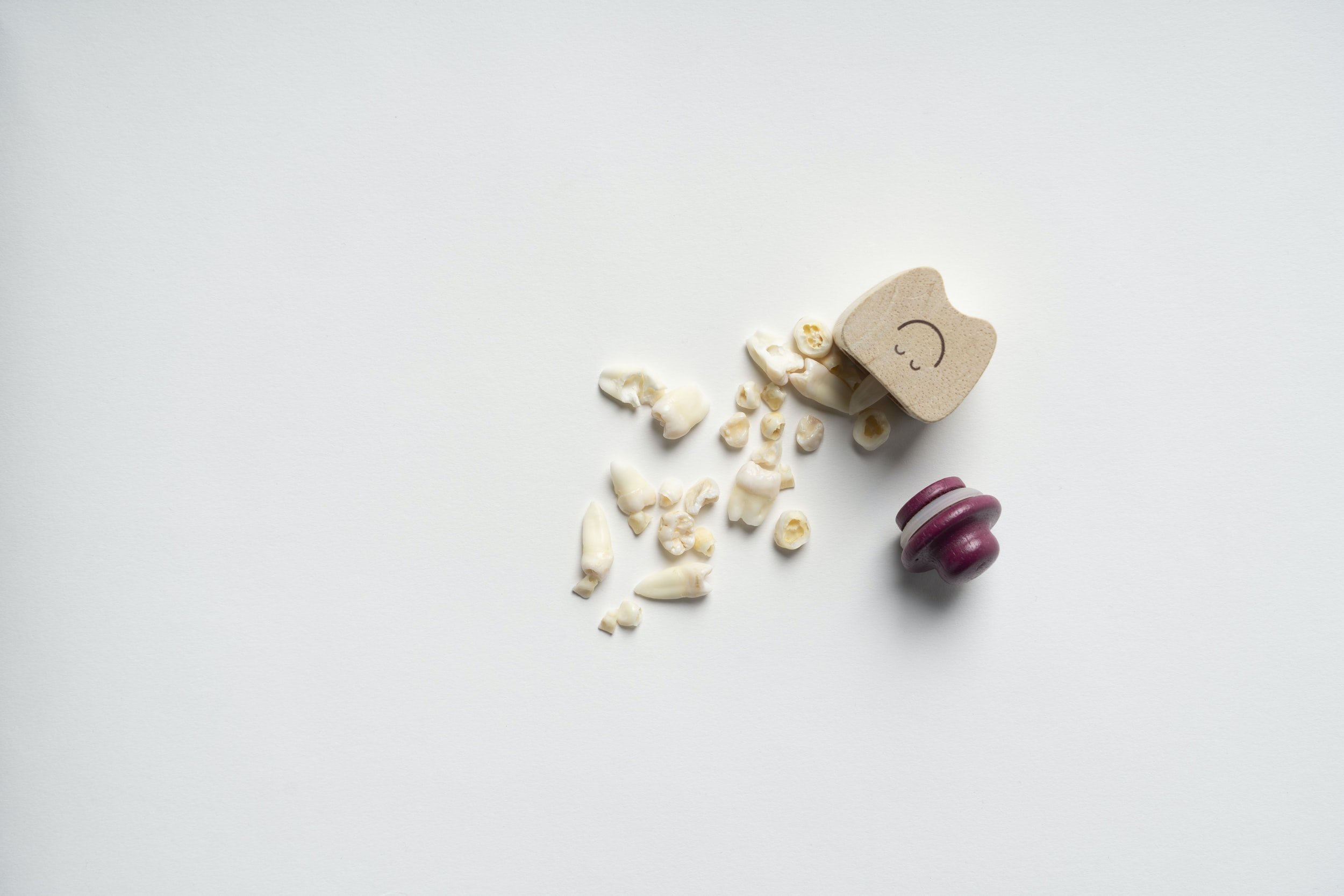
Pulling wisdom teeth can improve long-term taste function, research finds
Patients who had their wisdom teeth extracted had improved tasting abilities decades after having the surgery, according to a new study.
Patients who had their wisdom teeth extracted had improved tasting abilities decades after having the surgery, a new Penn Medicine study published in the journal Chemical Senses found. The findings challenge the notion that removal of wisdom teeth, known as third molars, only has the potential for negative effects on taste, and represent one of the first studies to analyze the long-term effects of extraction on taste.
"Prior studies have only pointed to adverse effects on taste after extraction and it has been generally believed that those effects dissipate over time," said senior author Richard L. Doty, PhD, director of the Smell and Taste Center at the University of Pennsylvania. "This new study shows us that taste function can actually slightly improve between the time patients have surgery and up to 20 years later. It's a surprising but fascinating finding that deserves further investigation to better understand why it's enhanced and what it may mean clinically."
Doty and co-author Dane Kim, a third-year student in the University of Pennsylvania School of Dental Medicine, evaluated data from 1,255 patients who had undergone a chemosensory evaluation at Penn's Smell and Taste Center over the course of 20 years. Among that group, 891 patients had received third molar extractions and 364 had not.
The "whole-mouth identification" test incorporates five different concentrations of sucrose, sodium chloride, citric acid, and caffeine. Each solution is sipped, swished in the mouth, and then spit out. Subjects then indicate whether the solution tastes sweet, salty, sour, or bitter.
The extraction group outperformed the control group for each of the four tastes, and in all cases, women outperformed men. The study suggests, for the first time, that people who have received extractions in the distant past experience, on average, an enhancement (typically a three to 10 percent improvement) in their ability to taste.
"The study strongly suggests that extraction of the third molar has a positive long-term, albeit subtle, effect on the function of the lingual taste pathways of some people," Kim said.
Two possibilities, the authors said, could explain the enhancement. First, extraction damage to the nerves that innervate the taste buds on the front of the mouth can release inhibition on nerves that supply the taste buds at the rear of the mouth, increasing whole-mouth sensitivity. Second, hypersensitivity after peripheral nerve injury from a surgery like an extraction has been well documented in other contexts. There is evidence, for example, from animal studies that repetitive light touch, which might occur during chewing, gradually accentuates neural responses from irritated tissue that can lead to progressive long-term tactile hypersensitivity. Whether this occurs for taste, however, is not known.
"Further studies are needed to determine the mechanism or mechanisms behind the extraction-related improvement in taste function," Doty said. "The effects are subtle but may provide insight into how long-term improvement in neural function can result from altering the environment in which nerves propagate."
The study was supported in part by the National Institute on Deafness and Other Communication Disorders (PO1 DC 00161).

Good toothbrushing habits in children linked to mother's wellbeing
Researchers have shown that postpartum depression can inhibit a mother's ability to instill healthy tooth brushing habits in children. The study demonstrates the need to foster greater mental support and management for mothers and incorporate these factors when assessing children's oral health.
A mother's mental health may play a larger role in a child's toothbrushing habits than previously thought.
It is no secret that brushing your teeth twice a day is highly effective in promoting healthy teeth and gums. The International Association of Pediatric Dentistry advocates brushing with toothpaste containing fluoride to prevent decayed, missing, or filled teeth -- known as childhood dental caries (ECC) -- in children.
Parents are instrumental in instilling good dental habits in their children.
In Japan, there is a worryingly high prevalence of ECC among children aged 3 years old. Postpartum depression and/or lack of affection caused by bonding disorders hamper a mother's ability to cultivate healthy dental practices in children, and researchers were keen to explore this link.
Dr Shinobu Tsuchiya from Tohoku University Hospital led a research group that analyzed approximately 80,000 mother-infant pairs from the Ministry of Environment's Japan Environment and Children's Study.
They found children with mothers suffering from postpartum depression or bonding disorders brushed their teeth less often. Likewise, the frequency with which children brushed their teeth increased when mothers showed strong affection towards their children.
The research group hopes their research will foster greater mental support and management for mothers and that doctors will incorporate these factors when assessing children's oral health.
"A mother's psychological well-being provides valuable screening information for identifying children at a high risk of ECC," said Tsuchiya
In future studies, Tsuchiya and her team hope to examine other environmental influences on poor oral health.

Baby teeth may one day help identify kids at risk for mental disorders later in life
The thickness of growth marks in primary (or 'baby') teeth may help identify children at risk for depression and other mental health disorders later in life, according to a ground-breaking investigation.
The team analysed 70 primary teeth collected from 70 children enrolled in the Children of the 90s study (also known as the Avon Longitudinal Study of Parents and Children) based at the University of Bristol. Parents donated primary teeth (specifically, the pointed teeth on each side of the front of the mouth known as canines) that naturally fell out of the mouths of children aged 5 to 7.
The results of this study could one day lead to the development of a much-needed tool for identifying children who have been exposed to early-life adversity, which is a risk factor for psychological problems, allowing them to be monitored and guided towards preventive treatments, if necessary.
The origin of this study traces back several years, when senior author Erin C. Dunn, ScD, MPH, learned about work in the field of anthropology that could help solve a longstanding problem in her own research. Dunn is a social and psychiatric epidemiologist and an investigator in MGH's Psychiatric and Neurodevelopmental Genetics Unit. She studies the effects of childhood adversity, which research suggests is responsible for up to one-third of all mental health disorders. Dunn is particularly interested in the timing of these adverse events and in uncovering whether there are sensitive periods during child development when exposure to adversity is particularly harmful. Yet Dunn notes that she and other scientists lack effective tools for measuring exposure to childhood adversity. Asking people (or their parents) about painful experiences in their early years is one method, but that's vulnerable to poor recall or reluctance to share difficult memories. "That's a hindrance for this field," says Dunn.
However, Dunn was intrigued to learn that anthropologists have long studied the teeth of people from past eras to learn about their lives. "Teeth create a permanent record of different kinds of life experiences," she says. Exposure to sources of physical stress, such as poor nutrition or disease, can affect the formation of dental enamel and result in pronounced growth lines within teeth, called stress lines, which are similar to the rings in a tree that mark its age. Just as the thickness of tree growth rings can vary based on the climate surrounding the tree as it forms, tooth growth lines can also vary based on the environment and experiences a child has in utero and shortly thereafter, the time when teeth are forming. Thicker stress lines are thought to indicate more stressful life conditions.
Dunn developed a hypothesis that the width of one variety in particular, called the neonatal line (NNL), might serve as an indicator of whether an infant's mother experienced high levels of psychological stress during pregnancy (when teeth are already forming) and in the early period following birth.
To test this hypothesis, Dunn and two co-lead authors -- postdoctoral research fellow Rebecca V. Mountain, PhD, and data analyst Yiwen Zhu, MS, who were both in the Psychiatric and Neurodevelopmental Genetics Unit at the time of the study -- led a team that analysed the teeth. The width of the NNL was measured using microscopes. Mothers completed questionnaires during and shortly after pregnancy that asked about four factors that are known to affect child development: stressful events in the prenatal period, maternal history of psychological problems, neighbourhood quality (whether the poverty level was high or it was unsafe, for instance), and level of social support.
Several clear patterns emerged. Children whose mothers had lifetime histories of severe depression or other psychiatric problems, as well as mothers who experienced depression or anxiety at 32 weeks of pregnancy, were more likely than other kids to have thicker NNLs. Meanwhile, children of mothers who received significant social support shortly after pregnancy tended to have thinner NNLs. These trends remained intact after the researchers controlled for other factors that are known to influence NNL width, including iron supplementation during pregnancy, gestational age (the time between conception and birth) and maternal obesity.
No one is certain what causes the NNL to form, says Dunn, but it's possible that a mother experiencing anxiety or depression may produce more cortisol, the "stress hormone," which interferes with the cells that create enamel. Systemic inflammation is another candidate, says Dunn, who hopes to study how the NNL forms. And if the findings of this research can be replicated in a larger study, she believes that the NNL and other tooth growth marks could be used in the future to identify children who have been exposed to early life adversity. "Then we can connect those kids to interventions," says Dunn, "so we can prevent the onset of mental health disorders, and do that as early on in the lifespan as we possibly can."
Dunn is also an associate professor of Psychiatry at Harvard Medical School. Mountain is now a postdoctoral research fellow at Maine Medical Center Research Institute. Zhu is now a doctoral student at the Harvard T.H. Chan School of Public Health.
Rare mineral from rocks found in mollusk teeth
Researchers discovered a rare mineral hidden inside the teeth of a chiton, a large mollusk found along rocky coastlines. Before this strange surprise, the iron mineral, called santabarbaraite, only had been documented in rocks.
Northwestern University researchers have, for the first time, discovered a rare mineral hidden inside the teeth of a chiton, a large mollusk found along rocky coastlines. Before this strange surprise, the iron mineral, called santabarbaraite, only had been documented in rocks.
The new finding helps understand how the whole chiton tooth -- not just the ultrahard, durable cusp -- is designed to endure chewing on rocks to feed. Based on minerals found in chiton teeth, the researchers developed a bio-inspired ink for 3D printing ultrahard, stiff and durable materials.
"This mineral has only been observed in geological specimens in very tiny amounts and has never before been seen in a biological context," said Northwestern's Derk Joester, the study's senior author. "It has high water content, which makes it strong with low density. We think this might toughen the teeth without adding a lot of weight."
The study will be published the week of May 31 in the Proceedings of the National Academy of Sciences.
Joester is an associate professor of materials science and engineering in Northwestern's McCormick School of Engineering. Linus Stegbauer, a former postdoctoral fellow in Joester's laboratory, is the paper's first author. At Northwestern during the research, Stegbauer is now a principal investigator at the Institute of Interfacial Process Engineering and Plasma Technology of the University of Stuttgart in Germany.
One of the hardest known materials in nature, chiton teeth are attached to a soft, flexible, tongue-like radula, which scrapes over rocks to collect algae and other food. Having long studied chiton teeth, Joester and his team most recently turned to Cryptochiton stelleri, a giant, reddish-brown chiton that is sometimes affectionately referred to as the "wandering meatloaf."
To examine a tooth from Cryptochiton stelleri, Joester's team collaborated with Ercan Alp, a senior scientist at Argonne National Laboratory's Advanced Photon Source, to use the facility's synchrotron Mössbauer spectroscopy as well as with Paul Smeets to use transmission electron microscopy at the Northwestern University Atomic and Nanoscale Characterization and Experiment (NUANCE) Center. They found santabarbaraite dispersed throughout the chiton's upper stylus, a long, hollow structure that connects the head of the tooth to the flexible radula membrane.
"The stylus is like the root of a human tooth, which connects the cusp of our tooth to our jaw," Joester said. "It's a tough material composed of extremely small nanoparticles in a fibrous matrix made of biomacromolecules, similar to bones in our body."
Joester's group challenged itself to recreate this material in an ink designed for 3D printing. Stegbauer developed a reactive ink comprising iron and phosphate ions mixed into a biopolymer derived from the chitin. Along with Shay Wallace, a Northwestern graduate student in Mark Hersam's laboratory, Stegbauer found that the ink printed well when mixed immediately before printing.
"As the nanoparticles form in the biopolymer, it gets stronger and more viscous. This mixture can then be easily used for printing. Subsequent drying in air leads to the hard and stiff final material," Joester said. Joester believes we can continue to learn from and develop materials inspired by the chiton's stylus, which connects ultra-hard teeth to a soft radula.
"We've been fascinated by the chiton for a long time," he said. "Mechanical structures are only as good as their weakest link, so it's interesting to learn how the chiton solves the engineering problem of how to connect its ultrahard tooth to a soft underlying structure. This remains a significant challenge in modern manufacturing, so we look to organisms like the chiton to understand how this is done in nature, which has had a couple hundred million years of lead time to develop."
The study, "Persistent polyamorphism in the chiton tooth: From a new biomaterial to inks for additive manufacturing," was supported by the National Science Foundation (award numbers DMR-1508399 and DMR-1905982), National Institutes of Health (award number NIH-DE026952), Air Force Research Laboratory (award number FA8650-15-2-5518) and Deutsche Forschungsgemeinschaft (award number STE2689/1-1).

A link between childhood stress and early molars
Research shows that children from lower-income backgrounds and those who go through greater adverse childhood experiences get their first permanent molars sooner. The findings align with a broader pattern of accelerated development often seen under conditions of early-life stress.
Early in her career neuroscientist Allyson Mackey began thinking about molars. As a researcher who studies brain development, she wanted to know whether when these teeth arrived might indicate early maturation in children.
"I've long been concerned that if kids grow up too fast, their brains will mature too fast and will lose plasticity at an earlier age. Then they'll go into school and have trouble learning at the same rate as their peers," says Mackey, an assistant professor in the Department of Psychology at Penn. "Of course, not every kid who experiences stress or [is] low income will show this pattern of accelerated development."
In the Proceedings of the National Academy of Sciences, Mackey, with doctoral student Cassidy McDermott and colleagues from Penn's School of Dental Medicine and the University of Missouri-Kansas City, shows that children from lower-income backgrounds and those who go through greater adverse childhood experiences get their first permanent molars earlier. The findings, generated initially from a small study and replicated using a nationally representative dataset, align with a broader pattern of accelerated development often seen under conditions of early-life stress.
"It's really important for us to understand how to detect early maturation sooner," Mackey says. "Right now, we're relying on seeing when kids hit puberty, which might be too late for some meaningful interventions. If we can inexpensively see that a child is experiencing this maturation earlier, we might be able to direct more intervention resources toward them."
A novel rating system
Broadly speaking, Mackey's lab studies how the brain changes and grows as people learn. It's well-established that stress during childhood speeds up maturation and that children who hit puberty earlier are at greater risk for both physical and mental health problems in adulthood.
Beyond that, in studies across primate species, molar eruption has been used to measure childhood length and correlates with a number of other developmental events. Similarly, for humans, the timing of dental events often plays a role in estimating biological age.
"That all made molar eruption a compelling developmental indicator," says McDermott, who is training to be a clinical psychologist.
It helped that more than 100 children, ages 4 to 7, had been participating in two Penn brain development studies, which included structural and functional MRI scans. "There's one type of MRI scan called a T2 weighted scan where you can visualize the morphology of the tooth pretty well," McDermott says. These scans -- typically used to look at the brain -- showed the researchers just how close these molars were to breaking through the gum line.
Once Mackey and McDermott realized this, they partnered with Katherine Hilton, then a student in Penn Dental Medicine, and Muralidhar Mupparapu, a professor in the Department of Oral Medicine, who developed a novel scale to precisely rate each tooth's position.
"The scale ranges from 1 to 4," McDermott says. "At the low end of the scale is 1, which is before the tooth has really developed at all. As the tooth emerges, there are intermediate stages, and the highest rating, a 4, is when the tooth is fully in the mouth and parallel with the other teeth." Four molars each received a score, which then got averaged, leaving a single score per individual.
Controlling for factors like age and gender, the researchers then looked for associations between early environment and molar eruption. "What we found is that income and adverse childhood experiences are both individually associated with molar eruptions status," McDermott says.
Replicating the findings
Those findings derived from just 117 participants, so although the correlation was clear, Mackey and McDermott hoped to replicate what they'd seen.
Collaborators at the University of Missouri-Kansas City told them about a large population-representative dataset called the National Health and Nutrition Examination Survey (NHANES), which is publicly available and includes dental data, demographic data, and family income, among other measures.
"Because our sample is only from one city and is much smaller than a population-representative study like that," McDermott says, "we saw it as an opportunity to verify that the findings exist outside of what we had collected in Philadelphia."
Though some facets differed -- NHANES measures dental development a little differently, for example -- the models showed similar results, indicating a connection between lower family income and earlier first molars.
Whether this overall trend is new or just now coming to light is something Mackey wants to study further. She's also curious about when the rate of maturation gets set. "Is it as early as in utero or is it dynamically adjusted based on stressors in the world?" she says. "If it's the latter, that tells you there are more opportunities to intervene."
Present implications, future work
There are still significant unknowns, as well as findings that need further examination, Mackey says. For example, the research team found racial disparities in this timing, with first molars emerging in Black children sooner than in white children.
"These race differences in molar eruption have been known for a long time, but no one thought critically about where they came from," she says. "It's consistent with higher levels of stress due to structural racism. This is a clear indication that it's not just speculation that experiences with racism can cause stress and early aging. They are having an effect on kids that we can't ignore."
For all children, a year-plus of pandemic-driven grief and social isolation most certainly amplified stress levels, making it even more important to understand who is at greatest risk for early maturation, Mackey says.
Yet she and McDermott emphasize that molar timing shouldn't become another parental fear. "What I really don't want is for parents to either worry or feel complacent just based on when their kids got their molars," says Mackey. "We don't have those data yet."
The Penn researchers are working on it. In the future, they hope to collaborate with dental offices to recruit children into studies based on their molar-eruption status. The goal would be to follow them into adulthood, to get more information on what precisely early first molars may indicate. "If this is the meaningful discovery that I think it is," Mackey says, "I would love for many scientists to jump on board and test these hypotheses."
Funding for this research came from the Jacobs Foundation, National Institute on Drug Abuse (Grant 1R34DA050297-01), and National Science Foundation.What would help, she thought, was a scalable, objective way -- a physical manifestation, of sorts -- to indicate how children embodied and responded to stresses in their world. Eruption timing of the first permanent molars proved to be just that.

Dental procedures during pandemic are no riskier than a drink of water, study finds
A new study's findings dispel the misconception that patients and providers are at high risk of catching COVID-19 at the dentist's office.
A new study's findings dispel the misconception that patients and providers are at high risk of catching COVID-19 at the dentist's office.
SARS-CoV-2 spreads mainly through respiratory droplets, and dental procedures are known to produce an abundance of aerosols -- leading to fears that flying saliva during a cleaning or a restorative procedure could make the dentist's chair a high-transmission location.
Ohio State University researchers set out to determine whether saliva is the main source of the spray, collecting samples from personnel, equipment and other surfaces reached by aerosols during a range of dental procedures.
By analyzing the genetic makeup of the organisms detected in those samples, the researchers determined that watery solution from irrigation tools, not saliva, was the main source of any bacteria or viruses present in the spatter and spurts from patients' mouths.
Even when low levels of the SARS-CoV-2 virus were detected in the saliva of asymptomatic patients, the aerosols generated during their procedures showed no signs of the coronavirus. In essence, from a microbial standpoint, the contents of the spray mirrored what was in the office environment.
"Getting your teeth cleaned does not increase your risk for COVID-19 infection any more than drinking a glass of water from the dentist's office does," said lead author Purnima Kumar, professor of periodontology at Ohio State.
"These findings should help us open up our practices, make ourselves feel safe about our environment and, for patients, get their oral and dental problems treated -- there is so much evidence emerging that if you have poor oral health, you are more susceptible to COVID," Kumar said.
The study was published Wednesday, May 12, in the Journal of Dental Research.
Previous research has shown that dental-procedure aerosols tend to land on providers' faces and the patient's chest, and can travel as far as 11 feet. But the studies, catching the spray in petri dishes placed on people, equipment and around the room, found only that bacteria existed -- they rarely identified the organisms and never determined where they came from. Saliva has been the presumptive source for a long time.
When saliva was considered potentially deadly at the start of the pandemic, Kumar decided a long-term answer was needed to settle the question of whether saliva is the source of dental aerosols.
For the study, the team enrolled 28 patients receiving dental implants and restorations using high-speed drills or ultrasonic scaling procedures in Ohio State's College of Dentistry between May 4 and July 10, 2020. Researchers collected samples of saliva and irrigant (the water-based cleaning solutions used to flush out the mouth) before each procedure and, 30 minutes after the procedure, aerosol remnants -- condensate -- from providers' face shields, the patient's bib and an area 6 feet away from the chair.
Kumar and colleagues then put genome sequencing technology to use that wasn't available in the petri-dish days. This allowed them to first characterize the microbial mix in pre-procedure saliva and irrigants, which they could then compare to organisms in the aerosol samples collected later.
With the analytical method they used, the researchers did not need to characterize the microbes -- they instead looked for variations in sequences that provided enough information to identify the family of bacteria or viruses to which they belonged.
"Some species that live in your mouth can closely resemble those in water and the environment. Using this method, we don't even have to know the names of these organisms -- you can tell whether they are exactly genetically identical or genetically different," Kumar said. "If you use this granular approach to see these very nuanced differences in the genetic code, you can very accurately identify where they're coming from."
No matter the procedure or where the condensate had landed, microbes from irrigants contributed to about 78% of the organisms in aerosols while saliva, if present, accounted for 0.1% to 1.2% of the microbes distributed around the room.
Salivary bacteria were detected in condensate from only eight cases and of those, five patients had not used a pre-procedural mouth rinse. The SARS-CoV-2 virus was identified in the saliva of 19 patients, but was undetectable in aerosols in any of the cases.
The findings are reassuring, but also make sense, Kumar said: Irrigant dilutes saliva -- a "thick, viscous" substance -- by an estimated 20- to 200-fold, and the research is validated by a 2020 study that reported a less than 1% COVID-19 positivity rate among dentists.
Kumar noted that dentistry has long been at the forefront of infection-control practices in health care. During the pandemic, new protocols have included strengthened ventilation systems, extra aerosol suction equipment, N95 masks and face shields on top of goggles, and extended downtime between patients. She is hopeful this study's findings will make practitioners and patients feel at ease about being in the dentist's office -- with continued stringent protection in place.
"Dental surgeons and hygienists are always at the forefront of the war against bacteria in the mouth, and they of course did not feel safe because they are front-line workers surrounded by aerosol," said Kumar, who has a periodontology practice of her own and was one of the procedure operators in the study.
"Hopefully this will set their mind at rest because when you do procedures, it is the water from the ultrasonic equipment that's causing bacteria to be there. It's not saliva. So the risk of spreading infection is not high," she said. "However, we should not lose sight of the fact that this virus spreads through aerosol, and speaking, coughing or sneezing in the dental office can still carry a high risk of disease transmission."
Co-authors of the study include Archana Meethil, Shwetha Saraswat and Shareef Dabdoub of Ohio State and Prem Prashant Chaudhary of the National Institute of Allergy and Infectious Diseases.

The Oral Microbiome Is Highly Site-Specific — And That Matters
Researchers have examined the human oral microbiome and discovered tremendous variability in bacterial subpopulations living in certain areas of the mouth. In many cases, the team was able to identify a handful of genes that might explain a particular bacterial group's habitat specificity.
Bacteria are not randomly distributed in the human body. In fact, many bacterial species show strong biogeography — meaning they thrive in very specific locations while being absent from others. This concept has major implications for understanding oral health, disease, and why “one-size-fits-all” approaches to probiotics or microbial therapies often fall short.
A recent study published in Genome Medicine and led by researchers at Harvard University provides new insight into just how specialized the oral microbiome really is.
What the Research Found
Using advanced sequencing and bioinformatic techniques, researchers examined bacterial populations from different areas of the mouth — including the tongue, cheeks, gums, and tooth surfaces.
Their findings were striking:
Bacteria of the same species can exist in genetically distinct forms depending on where they live in the mouth
Microbes found on the tongue are more similar to those on another person’s tongue than to bacteria elsewhere in the same mouth
Small differences in specific genes appear to help bacteria adapt to highly localized environments
In other words, the mouth is not a single ecosystem — it’s a collection of distinct microbial habitats, each with its own rules.
Why This Is Clinically Relevant
These findings help explain why:
Plaque bacteria behave differently than tongue bacteria
Certain oral diseases are site-specific
Simply “adding good bacteria” is unlikely to work unless those bacteria are adapted to the right environment
The study used a powerful approach called metapangenomics, allowing researchers to compare genetic variation across trillions of bacteria rather than relying only on a small number of lab-grown strains. This revealed meaningful differences between bacteria living naturally in the mouth and their laboratory counterparts.
Implications for Oral Health and Future Therapies
Understanding where specific bacteria thrive — and why — may eventually allow for more targeted strategies to manage oral disease. Rather than broadly altering the oral microbiome, future therapies could aim to:
remove harmful bacteria from specific sites
support beneficial microbes in the environments where they are most effective
better understand how disruptions in microbial balance contribute to disease
This research reinforces an important principle: oral health is deeply connected to biology at the microscopic level, and effective care requires respecting that complexity.
A Thoughtful Perspective
Studies like this don’t immediately change clinical treatment — but they deepen our understanding of how the oral environment functions and why precision matters. As oral medicine continues to evolve, insights into the microbiome will likely play an increasing role in prevention, diagnosis, and long-term care.
Reviewed and Updated January 2026