

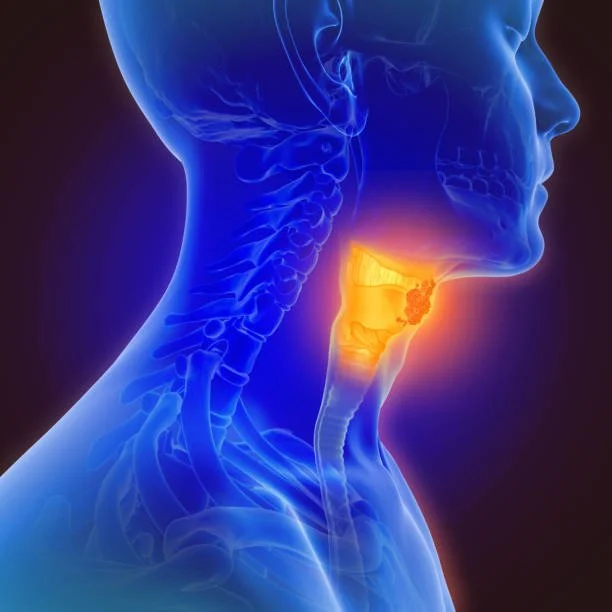
The 7 Warning Signs of Mouth Cancer: What You Need to Know
Oral cancer often develops quietly, appearing as nothing more than a stubborn sore or a faint patch of color. However, early detection is the single most important factor in successful treatment. From persistent mouth sores to subtle changes in your voice, recognizing these 7 warning signs can save your life. Learn what to look for during your monthly self-exam and when it’s time to seek a professional evaluation.
Mouth cancer (oral cancer) can be a silent traveler, often developing without significant pain in its early stages. However, your body usually provides subtle clues. At the office of CJ Henley DMD, we believe that being proactive is the best defense.
Recognizing these seven warning signs is the first step toward early diagnosis and successful treatment.
1. Persistent Sores That Don't Heal
A common red flag is a sore or ulcer that lingers for more than two weeks. While most canker sores clear up quickly, a persistent lesion—especially one that doesn't hurt—requires a professional oral medicine diagnosis to rule out malignancy.
2. Red or White Patches
Keep a close eye on the "landscape" of your mouth. Unusual red (erythroplakia) or white (leukoplakia) patches on the gums, tongue, or lining of the cheeks can be precancerous. If you notice a change in color that doesn't scrape off, it’s time for a check-up.
3. Unexplained Lumps or Thickening
If you feel a "bump in the night" (or day) inside your mouth or on your neck, pay attention. Any new lump or thickening of the tissues in the oral cavity or the head and neck region should be evaluated by a specialist immediately.
4. Chronic Sore Throat or Hoarseness
A sore throat that won't go away—or the constant feeling that something is "stuck" in your throat—can indicate issues deeper in the pharynx. Changes in your voice or persistent hoarseness are also signs that the vocal cords or surrounding tissues may be affected.
5. Difficulty Chewing or Swallowing
Pain or stiffness when moving your jaw or tongue can be more than just a muscle strain. If you find it increasingly difficult to chew, swallow, or speak, it may be due to a growth interfering with the normal function of your oral structures.
6. Numbness or Pain
Unexplained numbness in the tongue, lips, or face is a significant warning sign. This often happens when a tumor affects the underlying nerves. Similarly, persistent pain in the ear without any loss of hearing can sometimes be "referred pain" from the mouth.
7. Loose Teeth or Ill-Fitting Dentures
Unless you have advanced periodontal disease, teeth should stay put. If your teeth are loosening without an obvious cause, or if your dentures suddenly feel uncomfortable or "tight," it could indicate a change in the underlying bone structure of the jaw.
Why Early Detection Matters
The key to beating oral cancer is catching it before it spreads. During a routine exam, Dr. Henley performs thorough screenings to look for these signs, often identifying issues before they are visible to the naked eye.
Pro Tip: Perform a self-exam once a month in a well-lit mirror. Look under your tongue, check the roof of your mouth, and feel along your jawline for anything unusual.
If you have noticed any of these signs for more than 14 days, don't wait for your next cleaning. Early intervention is the most powerful tool we have.
Causes of a white tongue and how to get rid of it
White tongue is a term used to describe any area of the tongue that has a grayish-white coating on it. The coating may cover the entire tongue, or it may appear in patches.
A white tongue may be a surface coating or a change in the tissue itself. The appearance alone does not establish the cause. Persistent white patches, pain or a change that keeps returning should be examined.
Is a coating different from a white patch?
A coating can collect on the surface of the tongue. A white patch that remains in one area may need a different evaluation. Do not repeatedly scrape a sore area to test it; describe the change to your dentist instead.
Can dry mouth contribute?
Persistent dry mouth changes the oral environment and can increase the risk of fungal infection. Tell your dentist about medication changes, mouth breathing and any history of cancer treatment. Do not stop a prescribed medicine on your own.
Could it be thrush or oral lichen planus?
Oral thrush and oral lichen planus can cause white areas, but they need different treatment. A photograph or a description is not enough to choose an antifungal or another medication. Our oral medicine and diagnostic evaluation page explains the role of an examination.
What can I safely do at home?
Use a soft toothbrush and clean the tongue gently if it is comfortable. Avoid tobacco and anything that irritates the area. Persistent patches need evaluation even if oral hygiene is good; do not assume garlic, probiotics or vigorous scrubbing will treat the cause.
Does a white tongue mean cancer?
A white tongue does not by itself mean cancer. Some persistent tissue changes need further investigation. Oral cancer screening can identify an area that needs attention; a diagnosis may require additional testing.
Where can I have a tongue change checked in Jacksonville?
Contact C.J. Henley, DMD on Hendricks Avenue near San Marco. Note when the change began, whether it hurts and whether it has changed. Bring your medication list and relevant medical history.
Further reading: NHS: sore or white tongue and NIDCR: dry mouth.
Updated October 1, 2026.
Torus Palatinus: Causes, Cancer Risk, and When Treatment Is Needed
Learn what torus palatinus is, whether it is cancerous, when a hard bump needs a dental exam, and when removal may be recommended.
Oral health • CJ Henley, DMD • Jacksonville, Florida
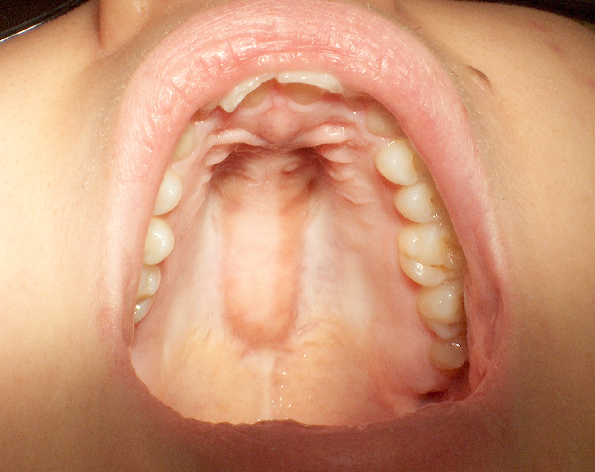
A torus palatinus is a benign bony growth in the middle of the roof of the mouth. It is usually painless and often needs no treatment.
Torus palatinus is not oral cancer. However, not every lump on the roof of the mouth is a torus. A dental examination can confirm the diagnosis, especially if the bump is newly noticed, changing, painful, bleeding, or associated with a sore that does not heal.
Quick answers
- What is it? An outgrowth of normal bone in the center of the hard palate, also called a palatal torus.
- Is it cancer? No. A confirmed torus palatinus is benign; an unexplained lump still needs an examination.
- Does it hurt? Usually not, although the thin tissue covering it can become irritated.
- Does it need removal? Usually not. Treatment is considered when it interferes with comfort, function, or a dental appliance.
What is torus palatinus?
A torus palatinus is a firm outgrowth of normal, mature bone along the midline of the hard palate—the bony front portion of the roof of your mouth. The plural is palatal tori. It usually develops slowly and is considered an anatomical variation rather than a disease.
Some tori form a smooth dome; others look like a broad ridge or a cluster of rounded lobes. They are more common in women. Estimates of how many people have them vary substantially between populations and studies. [1] [2]
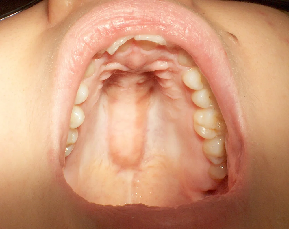

A typical palatal torus
- Centered on the hard palate
- Hard and bony
- Usually covered by intact tissue
- Stable or slowly growing
Changes to have checked
- Rapid enlargement or a new swelling
- Unexplained pain, bleeding, or numbness
- A sore that does not heal
- Persistent red or white patches
These features help explain what a dentist looks for. They are not a self-diagnosis checklist.
Is torus palatinus cancerous?
No. Torus palatinus is a benign bony growth, not oral cancer. The important step is confirming that a lump really is a torus. Other conditions can develop on the palate, and their appearances can overlap.
An oral cancer screening includes examining the mouth for concerning changes. If an area is suspicious, further testing or a biopsy may be needed; an examination alone cannot definitively rule out every condition.
When should a bump be examined?
- A newly noticed or changing lump, particularly one growing quickly.
- A swelling that is off-center, soft, or fluid-filled.
- A sore, ulcer, or red or white patch that persists for more than two weeks.
- Unexplained bleeding, numbness, or persistent pain.
These findings do not automatically mean cancer, but they deserve evaluation. Do not wait two weeks if symptoms are rapidly worsening. [4]
Seek urgent care for rapidly spreading swelling or difficulty breathing or swallowing.
Why an examination matters
A dentist considers the location, texture, history, surrounding teeth, and tissue over the bump. Our oral medicine evaluations help assess unusual lumps, sores, and other changes in the mouth.
| Condition | Possible features | Why evaluation matters |
|---|---|---|
| Palatal torus | A hard, usually slow-growing prominence in the center of the hard palate. | Often needs no treatment once the diagnosis is confirmed. |
| Salivary gland growth | A swelling that may occur toward the side or back of the palate; it can be painless. | Salivary gland tumors can be benign or malignant. A biopsy may be needed. |
| Dental abscess | Palatal swelling associated with an infected tooth; pain may occur. | Needs dental treatment to address the infection. |
| Nasopalatine duct cyst | A swelling near the front of the palate, sometimes in the midline. | A midline location alone does not establish that a lump is a torus. |
Further reading: National Cancer Institute: salivary gland tumors; diagnosis and management of nasopalatine duct cysts.
A typical torus can often be identified through examination. If the findings are unusual or surgery is planned, imaging may help assess the underlying anatomy. A CBCT scan provides a three-dimensional view when clinically indicated; it is not routinely needed for every torus. A biopsy is generally unnecessary for a typical torus but may be recommended when the diagnosis is uncertain.
Palatal and mandibular tori: a visual comparison
Palatal tori occur on the roof of the mouth. Mandibular tori occur on the inner surface of the lower jaw, beside the tongue. These photographs show examples in different people.


Tori vary in size and shape. Comparing your mouth with a photograph cannot confirm a diagnosis. Have a new or changing lump examined.
What causes torus palatinus?
The precise cause is not fully understood. Research points to a combination of inherited traits and environmental or functional influences. [3]
- Genetics: Tori can run in families.
- Bite forces: Teeth grinding and clenching, called bruxism, may contribute. They are not an established single cause of palatal tori.
- Other influences: Age, population differences, diet, and bone density have been studied, but their roles are not fully settled.
A palatal torus does not by itself prove that you grind your teeth or need a night guard.
Can torus palatinus cause pain or irritation?
Most palatal tori cause no discomfort. The tissue covering the bone is relatively thin, however, and hard or sharp foods can scratch it. Hot foods can burn the tissue, and an appliance that rubs the area can cause soreness.
A prominent torus may also interfere with the comfort or fit of an upper denture. Grooves between lobes can collect food, requiring attention during oral hygiene.
If the area becomes painful, have the cause assessed rather than assuming the torus explains every symptom. Avoid foods that repeatedly scrape the area, keep it gently clean, and have a rubbing appliance checked.
When is torus palatinus removal recommended?
Most people do not need removal. Surgery may be considered when the torus causes a specific problem: [1]
- It prevents a planned upper denture from fitting or functioning properly.
- It causes recurrent painful tissue injury that does not respond to conservative care.
- Its size interferes with speech, chewing, or swallowing.
The decision depends on your symptoms, anatomy, medical history, and dental treatment needs. A denture can sometimes be designed around a torus. Removal is not automatic simply because you need a denture.
What does removal involve?
When appropriate, an oral surgeon or another appropriately trained dentist lifts the overlying tissue, reduces and smooths the excess bone, and closes the area with sutures. Local anesthesia is used, with sedation considered according to the procedure and your needs.
A protective surgical stent may be used while the palate heals. Your surgeon will discuss risks such as bleeding, infection, wound-healing problems, and an opening into the nasal cavity. The nearby anatomy matters when planning surgery. [5]
Recovery varies with the size of the torus, the extent of surgery, and your health. Your dental team will provide instructions about food, cleaning, pain control, and follow-up. Initial comfort and complete tissue healing are not the same milestone.
Noticed a new bump on the roof of your mouth?
Schedule an examination with C.J. Henley, DMD at our Hendricks Avenue office in Jacksonville. We can assess the area and discuss whether observation, treatment, or a referral is appropriate.
3675 Hendricks Avenue, Jacksonville, FL 32207 · Contact and directions
Frequently asked questions
Does torus palatinus go away on its own?
A torus is made of bone and generally does not disappear on its own. If it causes no problems, it can usually be left alone and checked during routine dental visits.
Can a torus palatinus grow back after surgery?
Regrowth has been reported. Removal does not guarantee that a torus will never return. Your surgeon can discuss the likelihood in your situation and what to watch for afterward.
Can teeth grinding cause a bump on the roof of the mouth?
Grinding and clenching may contribute to the development of oral tori, but the relationship is not a proven single cause. An examination can assess both the bump and any evidence of grinding.
Does a palatal torus affect braces or clear aligners?
A torus does not necessarily prevent orthodontic treatment. Its size and position can affect appliances that contact the palate, such as some retainers and expanders. Your dentist or orthodontist will assess the fit and treatment plan.
Are bony bumps beside the tongue related?
Similar growths on the inner surface of the lower jaw are called mandibular tori. They often occur on both sides, beside the tongue. Like palatal tori, they are made of normal bone and usually need no treatment unless they cause problems. Not every lump in this area is a torus.
Can I remove or shrink a torus at home?
No home remedy has been shown to remove a palatal torus. Do not try to file, cut, or puncture it. If it bothers you, a dentist can determine whether treatment is needed.
About C.J. Henley, DMD
C.J. Henley, DMD is a general dentist in Jacksonville, Florida, whose practice includes cosmetic and restorative dentistry, oral medicine, and complex, medically involved dental care. He is a member of the Head & Neck Tumor Board at Baptist MD Anderson Cancer Center. Learn about his approach to dental care for patients with head and neck cancer.
References and further reading
- García-García AS, et al. Current status of the torus palatinus and torus mandibularis. Med Oral Patol Oral Cir Bucal. 2010;15(2):e353–e360.
- Haugen LK. Palatine and mandibular tori. A morphologic study in the current Norwegian population. Acta Odontol Scand. 1992;50(2):65–77.
- Loukas M, et al. The tori of the mouth and ear: a review. Clin Anat. 2013;26(8):953–960.
- National Institute of Dental and Craniofacial Research. Oral cancer: signs, symptoms, and diagnosis.
- Treatment of the Partially Edentulous Maxilla in the Presence of Tori. Dentistry Today. August 1, 2005.
- Torus palatinus: symptoms, causes and treatment. Cleveland Clinic.